Laser Eye Surgery for Presbyopia: A Comprehensive Guide

Laser Eye Surgery for Presbyopia: A Comprehensive Guide

As we age, our eyes undergo various changes, and one common condition that affects many individuals is presbyopia. This article aims to provide a comprehensive guide to laser eye surgery for presbyopia, focusing on understanding the condition, the basics of laser eye surgery, the procedure itself, and how to prepare for it.

Understanding Presbyopia

Presbyopia is a natural age-related vision condition that affects the ability to focus on near objects. It occurs due to the hardening of the lens and a decrease in its flexibility. This leads to difficulties in activities such as reading, using a smartphone, or seeing objects up close.

As we age, our bodies undergo various changes, and our eyes are no exception. The lens of the eye, which is responsible for focusing light onto the retina, gradually loses its elasticity. This loss of elasticity makes it more difficult for the lens to change shape and adjust its focus, resulting in presbyopia.

The Science Behind Presbyopia

To comprehend presbyopia, it’s essential to understand the underlying scientific principles. The eye’s lens, made of proteins and water, becomes less elastic over time. This results in a reduced ability to change shape, preventing the eye from properly focusing on close objects.

The hardening of the lens is caused by the gradual accumulation of proteins and other substances within its structure. These changes alter the lens’s refractive properties, making it less capable of bending light rays to form a clear image on the retina. As a result, near objects appear blurry, and individuals with presbyopia may find themselves straining their eyes or holding reading materials at arm’s length to see clearly.

Furthermore, the loss of flexibility in the lens affects its ability to accommodate, which is the process of adjusting the lens’s shape to focus on objects at different distances. Accommodation is crucial for clear vision, as it allows the eye to switch between near and far vision effortlessly. However, with presbyopia, the lens becomes less responsive to the muscles that control its shape, making it increasingly challenging to focus on close objects.

Symptoms and Diagnosis of Presbyopia

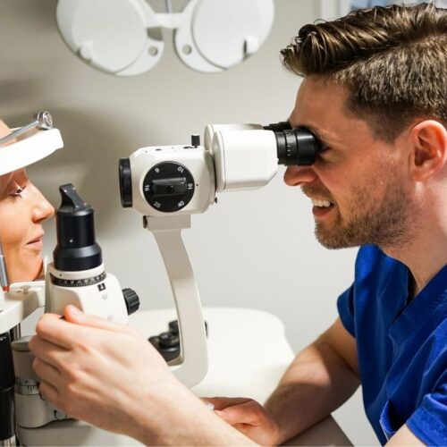
The common symptoms of presbyopia include blurred vision at a close distance, eye strain, and the need to hold reading materials at arm’s length. If you experience these symptoms, it’s crucial to undergo an eye examination with an optometrist or ophthalmologist for a proper diagnosis.

During an eye examination, the eye care professional will perform various tests to assess your visual acuity and determine the extent of your presbyopia. These tests may include a visual acuity test, where you read letters on a chart at different distances, and a refraction test, which measures the eye’s ability to focus light. Additionally, the eye care professional may use a device called a phoropter to determine the most suitable prescription for corrective lenses, such as reading glasses or multifocal lenses.

It’s important to note that presbyopia is a natural part of the aging process and affects nearly everyone to some degree. While it cannot be prevented or cured, there are several treatment options available to manage its symptoms and improve near vision. These options include wearing corrective lenses, such as reading glasses or contact lenses, undergoing refractive surgery, or using intraocular lenses.

By understanding the science behind presbyopia and recognizing its symptoms, individuals can seek appropriate vision care and make informed decisions about managing their near vision as they age.

Overview of Laser Eye Surgery

The best Laser eye surgery has revolutionized vision correction procedures, offering a safe and effective solution for various refractive errors. The surgery utilizes advanced laser technology to reshape the cornea, improving the eye’s ability to focus and reducing or eliminating the need for glasses or contact lenses.

Imagine waking up in the morning and being able to see the world clearly without reaching for your glasses or putting in your contact lenses. Laser eye surgery has made this a reality for millions of people around the world. By reshaping the cornea, the surgery corrects refractive errors such as nearsightedness, farsightedness, and astigmatism, allowing individuals to enjoy clear vision without the aid of visual aids.

But how does laser eye surgery work? Let’s delve into the basics of this remarkable procedure.

The Basics of Laser Eye Surgery

Laser eye surgery involves using a specialized laser to precisely reshape the cornea. The two most commonly performed procedures are LASIK (Laser-Assisted In Situ Keratomileusis) and SMILE (Small Incision Lenticule Extraction). Both procedures aim to correct refractive errors and improve vision.

During LASIK, a thin flap is created on the cornea using a microkeratome or a femtosecond laser. The flap is then lifted, and the underlying corneal tissue is reshaped using an excimer laser. Once the cornea is reshaped, the flap is repositioned, acting as a natural bandage.

On the other hand, SMILE is a minimally invasive procedure that involves creating a small incision in the cornea to access and remove a lenticule, which is a small piece of tissue causing the refractive error. This procedure requires no flap creation, making it a popular choice for individuals with thin corneas.

Both LASIK and SMILE offer quick recovery times and high success rates, making them preferred options for laser eye surgery.

Different Types of Laser Eye Surgery

There are several laser eye surgery options available, including LASIK, SMILE, PRK (Photo-Refractive Keratectomy), and LASEK (Laser-Assisted Sub-Epithelial Keratomileusis). Each procedure has its advantages and considerations, and the choice depends on factors such as corneal thickness, prescription, and personal preferences.

PRK is an alternative to LASIK and involves removing the thin outer layer of the cornea, known as the epithelium, before reshaping the cornea with an excimer laser. This procedure is suitable for individuals with thin corneas or those who may not be suitable candidates for LASIK.

LASEK combines elements of both LASIK and PRK. It involves creating a flap similar to LASIK but with a thinner layer of corneal tissue. The flap is then lifted, and the cornea is reshaped using an excimer laser. This procedure is often recommended for individuals with thin corneas or those at a higher risk of complications.

Choosing the right type of laser eye surgery is crucial for achieving optimal results. It is important to consult with a qualified ophthalmologist who can assess your specific needs and recommend the most suitable procedure. You can aslo read about Possible cataracts surgery risks and complications by visiting https://nwmedicalhypnosis.com/possible-cataracts-surgery-risks-and-complications/

Overall, laser eye surgery offers a life-changing solution for individuals seeking freedom from glasses or contact lenses. With advancements in technology and surgical techniques, more people can experience the joy of clear vision and improved quality of life.

Laser Eye Surgery for Presbyopia

Presbyopia, the age-related loss of near vision, has long been a challenge for individuals as they enter their 40s and beyond. While laser eye surgery has been primarily used to correct nearsightedness, farsightedness, and astigmatism, advancements in technology have paved the way for its usage in treating presbyopia as well.

Presbyopia occurs when the natural lens of the eye loses its flexibility, making it difficult to focus on close objects. This condition affects millions of people worldwide, leading to the need for reading glasses or bifocals. However, laser eye surgery offers a potential solution to this common problem.

How It Works

Laser eye surgery for presbyopia typically involves different techniques to improve near vision. These techniques may include creating a multifocal cornea or implanting multifocal or accommodative intraocular lenses.

One technique, known as monovision, involves correcting one eye for distance vision and the other for near vision. This allows the brain to adapt and merge the images from both eyes, providing a clear and balanced view of objects at various distances.

Another technique, called corneal inlays, involves inserting a small device into the cornea to improve near vision. These inlays can be made of various materials, such as hydrogel or synthetic polymers, and are designed to reshape the cornea, allowing for better focusing ability.

Benefits and Risks

The benefits of laser eye surgery for presbyopia are numerous. Patients often experience reduced dependence on reading glasses or bifocals, improved overall vision, and enhanced quality of life. Being able to see clearly without the constant need for corrective eyewear can greatly improve daily activities, such as reading, working on a computer, or enjoying hobbies.

However, like any surgical procedure, laser eye surgery for presbyopia does come with potential risks and complications. It’s important to discuss these with your surgeon before deciding to proceed with the surgery. Some common risks include dry eyes, glare or halos around lights, fluctuating vision, and the need for additional enhancements or touch-up procedures.

Additionally, not everyone is a suitable candidate for laser eye surgery for presbyopia. Factors such as the severity of presbyopia, overall eye health, and any existing eye conditions may affect the success and outcome of the procedure. A comprehensive evaluation by an experienced eye surgeon is necessary to determine if laser eye surgery is the right option for you.

In conclusion, laser eye surgery for presbyopia offers hope to those struggling with the loss of near vision. With advancements in technology and various surgical techniques, individuals can potentially regain their ability to see clearly at all distances, reducing the need for reading glasses or bifocals. However, it’s crucial to thoroughly discuss the benefits, risks, and suitability of the procedure with a qualified eye surgeon before making a decision. By clicking here you can read about Types of Eye Surgery for Refractive Errors.

Preparing for Laser Eye Surgery

Proper preparation is essential to ensure a successful outcome and a smooth recovery from laser eye surgery for presbyopia.

Initial Consultation and Evaluation

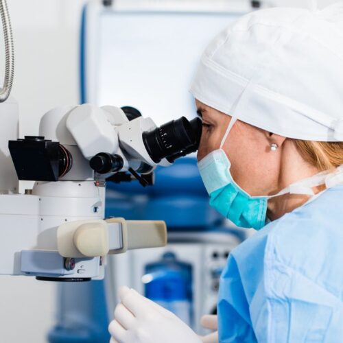
Your journey towards laser eye surgery begins with an initial consultation and a thorough evaluation of your eye health and visual needs. During this visit, your surgeon will assess your suitability for the procedure and address any questions or concerns you might have.

What to Expect Before Surgery

Prior to the surgery, you may need to undergo certain pre-operative procedures, such as refractive tests, corneal mapping, and measurements of your eye’s internal structures. Your surgeon will provide detailed instructions on how to prepare, including any necessary adjustments to your medication or lifestyle habits.

The Procedure: Step by Step

Understanding the sequence of events during laser eye surgery can help ease any apprehension you might have.

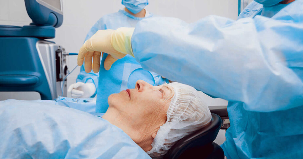
On the Day of Surgery

On the day of your surgery, you will be given specific instructions on what to do before arriving at the clinic or hospital. This may include avoiding eating or drinking beforehand, and you should arrange for transportation as your vision may be temporarily affected immediately after the procedure.
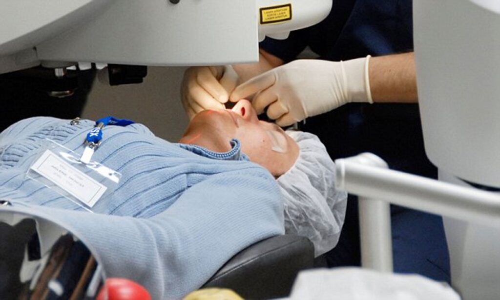
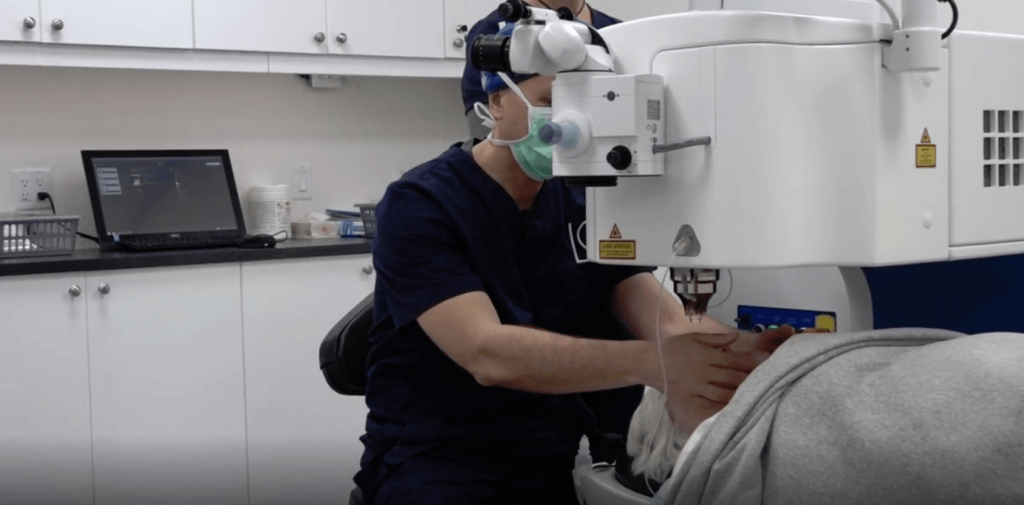
The Surgery Process

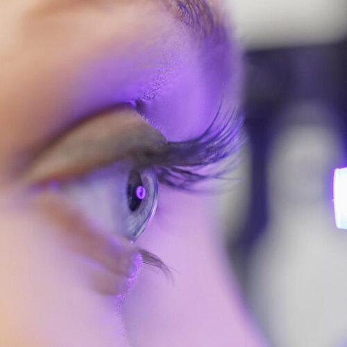
Laser eye surgery for presbyopia is typically a quick and painless procedure. After being given numbing eye drops, your surgeon will use the laser to reshape your cornea, following the predetermined plan discussed during your pre-operative evaluation. The surgeon will provide guidance and reassurance throughout the surgery.

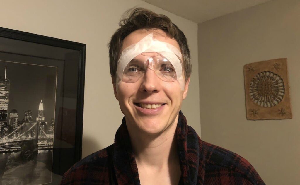
Following the conclusion of the surgery, you will rest for a short period and receive post-operative instructions and medications. It’s crucial to follow these instructions carefully to promote proper healing and minimize any potential complications.

Conclusion

In conclusion, laser eye surgery offers a comprehensive solution for individuals with presbyopia, enabling them to regain their near vision and reduce dependence on reading glasses or bifocals. By understanding presbyopia, the basics of laser eye surgery, and the preparation required, individuals can make informed decisions and embark on this life-changing journey with confidence.

Possible cataracts surgery risks and complications

Possible cataracts surgery risks and complications

Age-related alterations to our eyes are essentially unavoidable. For example, you could find that you now have trouble with glare, especially from headlights while driving at night, or that you require reading glasses to read a restaurant menu.

Cataracts, which more than half of Australians will have by the time they are 80 years old, often cause the latter issue. One of the research by personaleyes.com.au/cataracts shows when the lens, an eye component that concentrates light onto the retina to create a clear vision, begins to get clouded, a cataract develops.

According to Yale Medicine ophthalmologist Yvonne Wang, MD, a typical lens should be transparent, like to a windshield. “Your lens’ proteins begin to deteriorate when a cataract forms. The lens can no longer sustain that clarity when they alter in consistency.

See Also: Managing and preventing vitreous loss after cataracts surgery

A modification in the prescription for your glasses or contact lenses may be sufficient to assist when cataracts are initially developing. But since cataracts become worse with age, a lot of individuals end up having surgery to remove their current “cloudy” lens and replace it with a new artificial one. Thankfully, cataracts surgery is a routine and safe process.

Numerous improvements in cataracts surgery have also been made recently, including a laser-assisted procedure and a device called intraoperative aberrometry (ORA), which measures the eye further while the patient is under anesthesia. This makes it possible to measure the artificial lens used to replace the natural lens in cataracts surgery more precisely.

Possible cataracts surgery risks and complications

The options for lenses are also fast developing; they now go beyond just changing the lens. Some patients may no longer need glasses or contacts after cataracts surgery thanks to options like multifocal lenses, which not only repair the cataracts condition but also address vision issues.

Despite the fact that cataracts surgery is mostly safe, it can have certain risks. These may consist of:

Ocular cloudiness after cataracts surgery

A hazy layer known as posterior capsule opacification might develop behind your implant in 5 to 50% of instances. A YAG laser capsulotomy, an office-based procedure, may cure it in around 5 minutes.

Eyesight blurriness after cataracts surgery

Blurriness is typical in the days after cataracts surgery. Though sometimes it may take longer, it normally goes away in a few days.

Post-cataracts surgery floating

Floaters have the appearance of moving bits of dust. Floaters may not always need any special care, although they may indicate a retinal tear.

After cataracts surgery, dry eyes

After your operation, dry or gritty eyes are a frequent symptom. Eye drops are often used to treat this.

After cataracts surgery, eye discomfort

Serious discomfort might be an indication of an infection or other issues. To get the finest care, get in touch with your surgeon.

Following cataracts surgery, there is double vision

Double vision may be caused by a variety of things, but it often happens when your brain adjusts to your improved eyesight. Most likely, it will disappear in a few days.

Infection

Endophthalmitis is an infection of the fluids in your inner eye. Trusted Source It is estimated that 0.05 to 0.30 percent of cataracts operations will result in it.

Anaesthesia sensitivity

An allergic response is always a possibility after receiving anaesthesia. There are hardly many severe responses.

Cost of cataracts surgery

In the event that your operation is declared medically essential, insurance may pay for the costs. Location and operative method affect how much cataracts surgery costs.

The American Academy of Ophthalmology estimated in 2014 that out-of-pocket costs for this treatment might total about $2,500 when accounting for anesthesiologist, hospital, and surgeon expenses. This can imply that the price has increased. You can speak with local ophthalmologists to get a more precise figure.

Does Medicare pay for a cataract operation?

If cataracts surgery is performed using lasers or conventional surgical methods, Medicare will reimburse the expense.

A pair of corrective lenses or contacts that are required after surgery are covered to an extent of 80% by Medicare part B.

Possible cataracts surgery risks and complications

The prognosis and recovery after cataracts surgery

According to the National Eye Institute, cataracts surgery improves vision in roughly 90% of patients. The operation is thought to be secure and has the ability to restore the clarity of your vision that it had before the cataracts.

A second operation is necessary to address posterior capsule opacification, which occurs in 5 to 50% of patientsTrusted Source.

How long can cataracts surgery be put off?

Cataracts often grow slowly and are not seen as a medical emergency. The length of time you may safely put off having surgery depends on how quickly your cataracts are developing.

Although it’s normally better to get the operation as soon as possible, you should see a doctor to find out how long you may wait safely.

Cataracts will continue to worsen if ignored and might eventually cause total blindness.

It’s critical to identify and treat congenital cataracts in youngsters as soon as possible.

Takeaway

One of the most frequent and safest surgical procedures carried out in the US is cataracts surgery. The treatment takes just a few minutes and doesn’t need an overnight hospital stay. The cost is covered by Medicare and several insurance companies.

Ask your doctor whether you are a suitable candidate for surgery if you have cataracts that are impairing your eyesight.

Final thoughts

Cataracts surgery is one of the procedures that is done often. Over twenty million people throughout the world and over four million people in Australia get cataract surgery each year. These procedures are considered some of the safest that modern medicine has to offer. 

They have made it possible for millions of individuals to regain a higher quality of life via improvements in their vision. The cataracts experts at our eye clinic strive, in each and every clinical environment, to give our patients with the very best possible results.

When cataracts are first forming, a change in the prescription for your glasses or contact lenses may be enough to help. However, since cataracts worsen with aging, many people eventually need surgery to remove their present “cloudy” lens and replace it with a new artificial lens. Fortunately, cataracts surgery is a common and secure procedure.

Managing and preventing vitreous loss after cataracts surgery

Managing and preventing vitreous loss after cataracts surgery

Following cataracts surgery, impaired vision is associated with vitreous loss. Vitreous loss is less frequent among surgeons with expertise and those who perform a considerable number of cataracts operations. Techniques for classifying patients’ risks are available to help less experienced surgeons steer clear of high-risk patients. Before having cataracts surgery, patients must be counseled of potential risks and complications as part of the therapy for vitreous loss.

The vitreous body is shielded by the posterior lens capsule, an anatomical barrier, from forces associated with intraocular lens implantation, aspiration, and fragmentation of the lens. 4.4% of patients had capsule rupture and vitreous loss in the national cataracts surgery study conducted in the UK in 1997–1998. According to other studies, patient rates might range from 8.22 to 0.45 percent. A higher risk of vision-threatening effects such endophthalmitis, retinal detachment, and cystoid macular oedema is linked to vitreous degeneration. 2, 3, 4, 5, 6, 7 An even worse prognosis for those eyes is possible if the posterior capsule is disrupted and then lens fragments are displaced into the posterior segment.

Related: How to manage the long-term complications of cataracts surgery

It is crucial for the surgeon to refrain from actions that increase the risk of an eye catastrophe when vitreous loss occurs. These include non-vitrectomy techniques to collect lens fragments from the posterior area and phacoemulsification in the presence of vitreous.

The use of sutureless 23-gauge instruments and the pars plana route, as opposed to the anterior chamber, are benefits of doing anterior vitrectomies. There is an increased risk of retinal detachment, following glaucoma, and cystoid macular oedema with lens nuclear fragment displacement into the vitreous. It is advised that a retinal surgeon treat these eyes right away.

Managing and preventing vitreous loss after cataracts surgery

Prevention

The details of safe cataracts surgery are explored in more detail in recent literature, which is beyond the purview of this article.

The amount of vitreous loss is influenced by the surgeon’s expertise, surgical volume (the number of cataracts operations each surgeon does annually),12,13, the complexity of the cases, or case mix. Prior to cataracts surgery, scoring methods allow for a measurable estimation of risk in each case. By properly preparing patients, choosing the right anesthetic, and selecting the surgeon, problems may be anticipated and prevented. In high-risk eyes, Muhtaseb demonstrated that even seasoned consultants had an 8% vitreous loss rate and a 4% incidence of falling lens nuclei. It indicates that doctors experienced in vitrectomy and the removal of misplaced lens components from the posterior area are best suited to undertake cataracts surgery in such eyes.

Although the importance of the capsulorrhexis in the success of cataracts surgery has been appropriately emphasized17, the preparation of the patient, the anesthetic used, and the design of the incision may all influence the quality of the capsulorrhexis much earlier in the procedure. In high-risk eyes, such as those that have had vitrectomy18, problems may still occur even with perfect capsulorrhexis when an unstable anterior chamber depth causes anterior chamber depth changes, pupil constriction, and patient discomfort. 19 It’s important to comprehend how to take care of these eyes.

The care of the patient

Although it is not a common treatment, individuals now anticipate having cataracts surgery. The patient will have an easier time managing a significant intraoperative problem if informed consent included topics including predicted vitreous loss, lens matter displacement, and failure to implant an intraocular lens. This will reduce patient anxiety and help the patient and physician build trust before, during, and after cataracts surgery. 93.5 percent of patients want to be told if the risk is one in 50, and 62.4 percent want to be informed if the risk is one in 1000. Patients also want to be informed about rare effects. But recall accuracy of permission information is poor, especially for serious issues, making it difficult to adequately prepare patients for potentially hazardous operations.

Management: keeping a close eye

An internal alteration in the eye that is both subtle and sudden will signal the presence of vitreous loss. The sudden appearance of a red reflex altered lens nucleus mobility, excessive sideways displacement of the nucleus, abnormal movement of structures (like the pupil margin) remote from instruments in the anterior chamber, and traction transmitted through vitreous strands are just a few symptoms that may be seen.

Phacoemulsification should stop if it is still going on, and the probe should be withdrawn from the eye carefully to reduce strain on the vitreous. When the phacoemulsification probe is withdrawn, a viscoelastic substance may be injected into the anterior chamber to help stabilize any remaining lens fragments and avoid vitreous prolapse. 

Managing and preventing vitreous loss after cataracts surgery

The surgeon must then stop and meditate for a time. The vitrectomy equipment may be set up and, if required, a sub-anesthetic can be used while the situation is being evaluated. Tenon’s The lens nucleus or substantial portions of the lens material may be able to sink posteriorly if a reaction is delayed at this time. This is a problem that is controllable and often resolves well. When retrieving lens fragments using an anterior method, precipitation and force are harmful.

To finish cataracts surgery and securely implant an intraocular lens, vitreous loss management aims to remove all vitreous from the anterior chamber and operative site. Depending on the intricacy of the case and the main surgeon’s skill level, any, all, or none of these activities may be completed by the primary surgeon. A vitreous cutter and a separate infusion provided by an anterior chamber maintainer should be used to remove vitreous. Because it produces a flow conflict between infusion and aspiration and makes it difficult to maintain control of intraocular pressure, the use of a coaxial infusion sleeve around a vitreous cutter is not advised.

A drawback of so-called “dry” vitrectomy techniques is their inability to maintain a constant intraocular pressure. The cutter should be placed into the pars plana, either utilizing an anterior chamber or pars plana infusion, to collect vitreous from the anterior chamber. The vitreous may now be removed from deep beyond the posterior capsule, which is more challenging and dangerous when the cutter is put via the anterior chamber. This also decreases anterior chamber manipulation and vitreous incarceration in the corneal incision. Although triamcinolone particles may be used, their use is not necessary to view vitreous strands in the anterior chamber.

The procedure used a 25-gauge needle for sutureless vitrectomy. The bigger 23-gauge sutureless devices provide more control and could be more successful in removing vitreous as well as retained lens debris. Soft lens debris may easily be removed using the vitrector. 

The nuclear material may either be emulsified and aspirated by reintroducing the phacoemulsification probe or intact nuclear fragments can be removed through an enlarged incision once the anterior chamber has been completely cleared of vitreous. Nuclear fragments may be stabilized with viscoelastics or a lens glide29 before to removal from the anterior chamber.

Forceful efforts to remove nuclear fragments without a pars plana vitrectomy may cause significant retinal tears and detachment if they are moved posteriorly past the plane of the posterior capsule.

How to manage the long-term complications of cataracts surgery

How to manage the long-term complications of cataracts surgery

Phacoemulsification cataracts surgery is quickly replacing traditional cataracts treatments because it is a safe procedure with a low risk of complications. After a brief, painless outpatient procedure, the vast majority of patients report significant improvement in their vision. 

Although complications after cataracts surgery are incredibly rare, they can happen and harm the patient’s vision. Since many of these problems become apparent the day after cataracts surgery, it is advised that patients be examined the following day. Any issues that are found can then be dealt with quickly, enabling appropriate healing and recovery to occur.

The surgical trauma, particularly the ultrasonic energy and the fluid flowing into the anterior chamber, can result in ocular edema (blurred vision). The vision will be obstructed while the endothelial cells are working to remove the water from the corneal stroma. 

Although corneal edema is less common due to the use of high-quality viscoelastic materials, prolonged cataracts surgery on a dense nucleus can still cause localized or, in some cases, widespread corneal swelling. A hypertonic saline solution and steroid-based inflammation management can help hasten the recovery of vision.

How to manage the long-term complications of cataracts surgery

An elevated intraocular pressure (IOP)

The IOP should be within physiologic bounds for most patients. Depending on whether an incision has been made, it could be higher or lower due to residual viscoelastic. It is possible to use topical and oral medications to treat high intraocular pressure (IOP), but the patient needs to be kept under close observation. When the viscoelastic is removed from the anterior chamber, pressure usually returns to normal. Because tapping the anterior chamber to release aqueous is often just a temporary fix and exposes the eye to a high risk of infection, it is not generally recommended.

As there is usually some degree of inflammation brought on by surgical operations, it is normal to see some anterior chamber cells and flare the day after cataracts surgery. It is not normal to have an abnormally strong anterior chamber response to fibrin or a hypopyon; this might be a sign of endophthalmitis or the inflammation-causing condition toxic anterior shock syndrome. 

Both need routine steroid administration to keep the inflammation in check, but the latter requires a diagnostic tap, an antibiotic injection, and prompt treatment to prevent eyesight and the eye from being irreparably damaged. Even though endophthalmitis usually doesn’t show up until several days following cataracts surgery, it may happen as early as the first postoperative day.

Anatomic problems are a symptom that surgery is necessary.

Tiny remnants of a cataracts may remain in the anterior chamber after phacoemulsification. This happens as a result of the cataracts being divided up after surgery and a possible little component remaining in the anterior chamber. These pieces of the cataracts have the potential to cause serious regional corneal damage and inflammation. A tiny, wispy cortical piece may be treated medically with steroids and a focused YAG laser treatment to break it up into smaller fragments and increase the surface area to volume ratio, but larger pieces will need surgical intervention. In cases of cataracts surgery when there has been a posterior capsule rupture and there are still lens pieces in the vitreous, a pars plana lensectomy and vitrectomy are indicated. In instances requiring cataracts surgery, a referral to an accomplished vitreoretinal specialist is advised.

How to manage the long-term complications of cataracts surgery

IOL subluxation: If the IOL implant is not securely fastened, it runs the risk of slipping out of place and being subluxated. This is more often seen in situations where the lens was implanted into the ciliary sulcus as a consequence of a flaw in the eyeball’s posterior capsule. While minor episodes of IOL subluxation are conceivable, a significant displacement of the IOL from the visual axis has to be surgically corrected. In certain circumstances, this kind of suture attachment may assist in ensuring the IOL’s long-term stability and centering.

Leaking from an incision: If the IOP is low, it’s likely that there is leaking from an incision. A fluorescein dye leakage test may help identify the leak’s cause and determine how much fluid is escaping. Significant leakage from an incision indicates that a suture will very probably be required to close the wound, prevent further hypotony, and lower the risk of infection. The presence of iris tissue trapped within an incision is a sign that there is a problem with the incision since the iris often functions as a stopper to help shut leaky incisions.

Issues that linger for a long period

While many potential complications after cataracts surgery may be found on the first postoperative day, others take longer to appear. Following cataracts surgery, retinal issues such cystoid macular edema or retinal fractures are frequent and may appear days or weeks later. Only when the capsular bag has closed and the IOL has been positioned in its final position can relative refractive errors be accurately evaluated. Due to its frequency and tendency to appear weeks or months after cataracts surgery, posterior capsule opacification may not even be considered a side effect of the treatment.

Modern cataracts surgery is a wonderful procedure, but an ophthalmologist must still be involved both during and after the procedure. In the end, the surgical procedure and the healing process that takes place in the postoperative period together define the patient’s ultimate visual outcome.

Final thoughts

This article will keep you on track as far as making the appropriate decisions throughout your cataracts surgery recovery process if you’re wanting to manage the long-term problems of cataracts surgery.

Cataracts surgery is one of the most often used therapies. Each year, more than 4 million Americans and over 20 million other people get cataract surgery. One of modern medicine’s safest procedures is this one. Millions of individuals are able to regain a higher quality of life because to their better vision. Our eye clinic’s cataract experts always strive to provide patients the finest results possible in every clinical situation.

Looking to get professional advice on cataracts surgery? We’re only a message away.